Demystifying Misconceptions about Infertility and conception using ART

World Health Organization (WHO) defines Infertility as Failure to achieve pregnancy after 12 months or more of regular unprotected sexual intercourse.

Here are some of the misconceptions about Infertility.

Myth 1: Treatment for Infertility is complex and expensive
It depends upon the condition of the patient. Depending upon the degree of the complexity of the situation different procedures like Ovulation Induction (OI), Intra Uterine Insemination (IUI), and Invitro fertilization (IVF) are available. The procedures need not be complex and expensive. Except for the case of IVF, others are out-patient treatments

Myth 2: Infertility is due to a female’s inability
On the contrary, males contribute significantly to infertility[1].

Myth 3: Infertility treatment assures success
Depending upon several aspects the success rate is estimated to be about 40% or more[2]

Myth 4: Infertility treatment is affordable only to the Rich
Indian medical eco-system is making conscious efforts in ensuring that the treatment is accessible and affordable without compromising on quality.

Myth 5: Infertility treatment can be taken at any age
While it seems to be true age plays a significant role in achieving success. The odds of success improve if one takes it at an earlier age.[3]

Let’s collaborate…

If you have questions or if you want to be a part of this myth-busting venture about infertility and conception using ART, you can reach out to contact@spovum.com or send a message on 9513646022

“If you or anyone you know need help in and around Bagalkote reach out to Mane Hospital’s LifeNu IVF Center. It is quite accessible and affordable. It is situated very close to both the Railway station as well the Bus Stand of Bagalkote. LifeNu in collaboration with SpOvum intends to bring the best in class ART technology to its patients!”

– LifeNu IVF Center, Mane Hospital

References:

  1. Kumar N, Singh AK. Trends of male factor infertility, an important cause of infertility: A review of literature. J Hum Reprod Sci. 2015 Oct-Dec;8(4):191-6. doi: 10.4103/0974-1208.170370. PMID: 26752853; PMCID: PMC4691969.https://pubmed.ncbi.nlm.nih.gov/26752853/
  2. Malhotra N, Shah D, Pai R, Pai HD, Bankar M. Assisted reproductive technology in India: A 3 year retrospective data analysis. J Hum Reprod Sci. 2013 Oct;6(4):235-40. doi: 10.4103/0974-1208.126286. PMID: 24672161; PMCID: PMC3963305.https://academic.oup.com/hropen/article/2017/2/hox011/4062213
  3. Megan L. Sneed, Meike L. Uhler, H. Edward Grotjan, John J. Rapisarda, Kevin J. Lederer, Angeline N. Beltsos, Body mass index: impact on IVF success appears age-related , Human Reproduction, Volume 23, Issue 8, 1 August 2008, Pages 1835–1839, https://doi.org/10.1093/humrep/den188


SpOvum at LifeNu

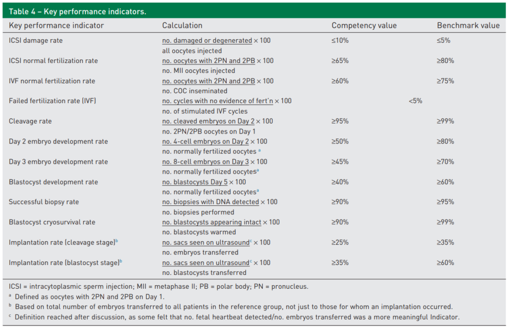
SpOvum has been operating successfully at Mane hospital’s LifeNU. While SpOvum is taking care of their operations including Technology-enablement, Embryologists, Dashboard, Maintenance, Calibration ad a bit of counseling. Recently SpOvum has also installed its flagship product- RoboICSI® holder, and smART Alerts system. SpOvum is making a conscious effort to see that LifeNU meets its KPIs along the Vienna consensus.

Largely, it has been successful in achieving so. For example, we have got an ICSI Damage rate of 0% (Benchmark from Vienna consensus is ≤ 5%), a Day3 Embryo Development rate of 84% (Benchmark from Vienna consensus is ≥ 70%), Blastocyst cryo survival rate of 100% (Benchmark from Vienna consensus is ≥ 99%)

SpOvum is making strides in ensuring IVF hospitals excel in providing quality IVF treatment to patients.

SpOvum’s brief interaction with the Oasis team

SpOvum® got an opportunity to present its product portfolio to the team of Oasis fertility headed by Dr Krishna Chaitanya in the early morning of 29th November 2022. Though it was originally expected to be brief (about fifteen minutes) it got extended! Thanks to the intriguing interaction. Dr Krishna and Dr Gaurav had some wonderful questions and were appreciative of SpOvum’s initiative in bringing innovations to the A.R.T. space through Robotics, Automation, and AI.

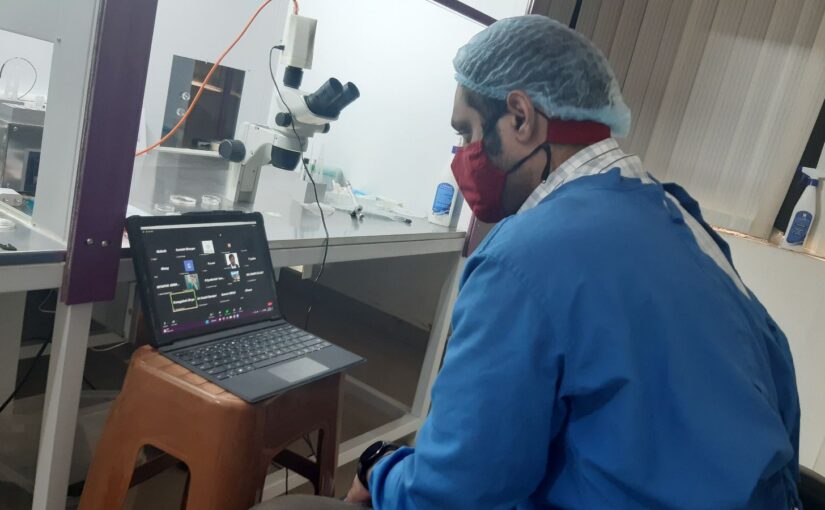
SpOvum presented its flagship product – RoboICSI® and its ICSI workbench – RIWB. The scope of automation and some of the key aspects that the innovation can address were discussed. The meeting happened virtually over the Zoom video communication platform. Further, SpOvum presented a live demo of the RoboICSI® gripper-holder, the workbench comprising the programmable manipulators, the Electronic aspiration system controlled by a click-based controller, and an indigenously designed button controller.

SpOvum wishes to engage with the Oasis fertility team to bring a synergetic approach to addressing some of the needs of the IVF ecosystem. As the vision statement of Oasis indicates “To be the trusted experts and leaders in providing evidence-based fertility treatments with compassion” SpOvum has found a common ground for a strong collaboration. Looking forward to a brighter future!


Beauty lies in the eye of the beholder

The connection of beauty to the eyes of the beholder is much deeper than what it looks. Different people have different perception of reality or on things. Some might find somethings good while other may find it bad. In this study let us concentrate on Morphological inspection.It is the best tool and it has wide range of observations for a given sample…Let’s find out how?. The image below was asked for assessment to the embryologists in a conference and was told to grade the oocyte accordingly.

Oocyte image given for the quiz [courtesy : ESHRE Atlas of human embryology]


Around 20 of them took up the quick quiz. The image that was given during the quiz was taken from ESHRE Atlas of human embryology. Below the image were a list of buttons which were related to the grading of the oocytes like: Maturity, Size, Zona, SER etc. They had to select the appropriate options referring to the image.

The result of the quiz was made into a pie charts for better representation and understanding. We were surprised to see the answers obtained.

Zona appearance, for example appeared to be Uniform for 63.2% of them and Non-uniform for 36.8%.

Likewise, maturity of oocyte for a M2 oocyte was 83.3%, M1- 11.1% and GV 5.6%.

Maturity graph [M2;GV;M1]

Grading the oocyte based on quality, 50% has told it is good, 25% abnormal, 15% approximately mature and 10% said almost normal.

Grade quality of the oocyte

The polar body had sections like – Shape, texture, size and fragments. Shape: 2 of them found it to be irregular, 10 of them opted oval and 5 of them found the oocyte to be round. Based on the texture of PB- 10 of them found it smooth and 7 of them rough. The PB size was selected normal by 15 of them, smaller and enlarged by 5 of them. 7 of them found the PB to be fragmented whereas 9 of them found the PB to be normal without any fragments.

Polar body grading


Zona pellucida grading of the oocyte

Vacuoles and SER have got the same percentage of answers. Absent is 62.5% and Present is 37.5%.

Vacuole and SER grading of the oocyte


SpOvum Platform is adopted at DYU LittleBaby Healthcare, Bengaluru

SpOvum team is delighted to associate with the DYU LittleBaby Clinic at HSR Layout, Bengaluru.

We also are delighted that this will be the First Center in the World to adopt the entire SmART IVF platform which SpOvum offers.

You can read more about the clinic here: https://child-development-center-bangalore.business.site

SpOvum's SmART-IVF platform is adopted at DYU LittleBaby center

Team SpOvum wishes them a great success in the days to come …

Bio-Insights- Sperms

Structure of Spermatozoon 

A morphologically normal sperm cell is about 40-50 µm in length and consists of a head and tail. [2]

Head: It mainly consists of nucleus and acrosome. Sperm head performs two functions- genetic and activation. The genetic function is embodied in the sperm nucleus which consists of DNA and nuclear proteins and thus is responsible for the transmission of hereditary characters from the male. The major part of the sperm head is occupied by the nucleus about 65%, which determines the sperm head shape. The sperm head anterior end is covered by a cap-like structure called acrosome. The acrosome is represented by Golgi complex and it contains a number of hydrolytic enzymes, such as hyaluronidase and acrosin, which are required for fertilization. During fertilization, the acrosomal membrane fuses with the oocyte cytoplasmic membrane and followed by acrosomal reaction, an event where the acrosomal enzyme is released from the head tip. Sperm head measures between 4.0-5.5 µm in length and 2.5-3.5 µm in width. [1 and 2]

Structure of a Sperm

Neck: It is a short, slightly constricted segment made up of projections located between the head and the tail portion. Neck differs clearly from the head and also from the rest part of the tail. [1 and 2]

Tail: The tail measures 40-50 µm in length and provides motility for the cell. Sperm cell’s entire motility apparatus is contained in the tail. The tail can be divided into the mid-piece (anterior portion), principle piece, and end-piece (posterior portion). Mid-piece supports the head at exactly the center position. The mid-piece consists of tightly packed mitochondria surrounded by a sheath. The mitochondria in the mid-piece supply energy in the form of ATP for tail movement. The principle piece is the longest part of the tail and comprises most of the propellant machinery. Motility plays the main role in sperm transport through the cervix. [2]


Abnormal Sperm Morphology

Teratospermia is a condition characterized by the presence of sperm with an abnormal morphology that affects fertility in males.  Normal sperm exhibits an oval-shaped head with a regular outline and a cap (acrosome) covering more than one-third of the head surface. The mid-piece is slender, less than one-third of the width of the head, straight and regular in outline. The tail is slender, uncoiled and should present a regular outline. Abnormal sperm morphology is classified as a defect in the head, midpiece or tail of the sperm.

Sperm Head Abnormalities

Head defects include large, small, tapered, pyriform, round, amorphous heads, heads with a small cap area and double heads, as well as any combination of these. Globozoospermia, where the sperm head appears small and round due to the failure of the acrosome to develop is one of the examples of a head defect.

Mid-piece defects include “bent” neck (where the neck and tail form an angle greater than 90% to the long axis of the head), thick/irregular mid-piece, abnormal thin mid-piece, as well as any combination of these.

Sperm Tail Abnormalities

Tail defects Include short, multiple, hairpin, broken or bent (>900) tails, tails of irregular width, coiled tails, as well as any combination of these.


Sperm Defect

Possible related observations Possible associated functional anomaly

Elongated head

Abnormally shaped head and abnormally condensed chromatin Immature chromatin/fragmented DNA/increased aneuploidy

Thin head

Abnormally shaped head and abnormally condensed chromatin

Immature chromatin/fragmented DNA

Microcephalous head

Excessive shrinkage of the nucleus and abnormally condensed chromatin

Immature chromatin/fragmented DNA

Macrocephalous head

Insufficient shrinkage of the nucleus and abnormally condensed chromatin

Increased aneuploidy

Multiple heads

Two or more closed or dissociated heads with or without a common acrosome or midpiece

Handicaps migration through mucus and oocyte vestments/fragmented DNA

Abnormal acrosome region

Absent or abnormally shaped or sized acrosome, incomplete acrosome and/or abnormal appearance of the underlying nucleus

Abnormal acrosome reaction/fragmented DNA

Thin midpiece

Partial/absent mitochondrial sheath

No or reduced ATP available for cell propelling

Bent tail

Misaligned midpiece and head or sharply bent midpiece/tail

Impairment of syngamy and cleavage, abnormal cell propelling, handicaps migration through mucus and oocyte vestments

Absent tail

Various anomalies of the neck region

Fragility of the neck structure and sperm moving forward is not possible

Short tail

Abnormally shaped periaxonemal and sometimes axonemal structures/dysplasia of the fibrous sheath

Immotility or severe dyskinesia


Irregularly shaped tail


Abnormally shaped periaxoneamal and sometimes axonemal structures/dysplasia of the fibrous sheath

Abnormal motion

Coiled tail

Completely or partially coiled tail often within a huge cytoplasmic remnant

Sperm moving forward is not possible

Multiple tails more than one tail

Partially dissociated tails connected to a single or to multiple heads or tails knitted together over a variable length

Abnormal motion: handicaps migration through mucus oocyte vestments


Semen Analysis

This is an evaluation of a certain characteristic of male’s semen and the sperm. This helps in evaluating male fertility

  • Color: Normally semen appears in whitish-gray in color. As the man ages, it appears as a yellowish tint. Presence of blood in semen (hematospermia) a rare condition which results in brownish or red color ejaculate. Deep yellow or greenish color appearance of semen is due to medication. Other causes of unusual semen color are due to STI’s, genital surgery and injury to male sex organs.
  • Volume: Semen volumes between 2.0 ml and 5.0 ml are considered to be normal. WHO regards 1.5 ml as the lower reference limit.
  • pH: According to WHO the normal semen pH is in the range of 7.2-8.2. An acidic pH ejaculate indicates one or both of the seminal vesicles are blocked. A basic pH ejaculate indicates an infection. pH value outside of the normal range is harmful to sperm and affects their ability to penetrate the egg.
  • Viscosity: It measures seminal fluid’s resistance to flow. High viscosity may interfere with the determination of sperm motility, concentration and antibody of spermatozoa. Normally semen coagulates upon ejaculation and usually liquefies within 15-20 min. Liquefaction time within 60 min is considered as the normal range.
  • Motility: The efficient passage of spermatozoa through the cervical mucus is dependent on rapid progressive motility, that is, spermatozoa with a forward progression of at least 25 µm/s. Reduced sperm motility can be a symptom of a disorder related to male accessory sex gland secretion.

>Rapid progressive motility- Moves at >25 µm/s at 370C and >20 µm/s at 200C

>Non-progressive motility- Move at <5 µm/s

>Immotility

Reference:

  1. http://www.biologydiscussion.com/notes/structure-functions-and-types-of-mature-sperm-in-animals-biology/768
  2. https://www.researchgate.net/publication/283864069_Sperm_Biology_from_Production_to_Ejaculation
  3. file:///C:/Users/BBC_common/Downloads/fertilitypedia-abnormal-sperm-morphology%20(1).pdf
  4. https://www.ncbi.nlm.nih.gov/pmc/articles/PMC3114587/
  5. https://en.wikipedia.org/wiki/Semen_analysis


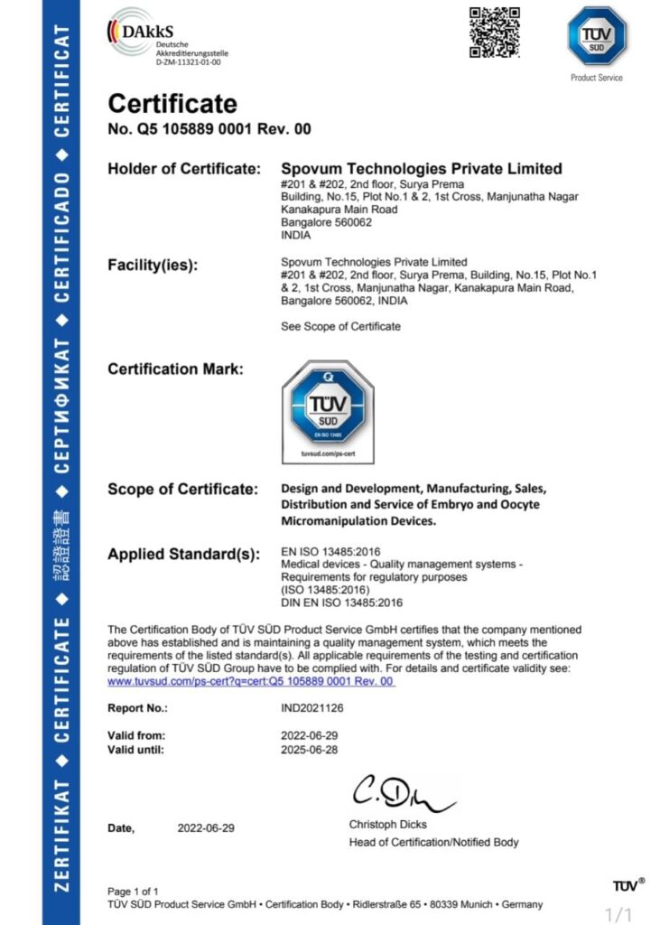
SpOvum is now ISO 13485 certified

SpOvum has received the ISO 13485 certificate. This certificate was issued by TUV SUD.

SpOvum ISO 13485 certificate

What you should know regarding ISO 13485
What is a medical device?
A product that is designed for use in the diagnosis, prevention, and treatment of illnesses or other medical disorders is known as a medical device. Examples include instruments, machines, implants, and in vitro reagents.

Whom does ISO 13485 serve?
Organizations engaged in the design, production, installation, and maintenance of medical devices and related services are intended users of ISO 13485. Additionally, it can be utilized by internal and outside parties, including certification organizations, to support their auditing procedures.

Compliance with ISO 13485
Like other ISO management system standards, ISO 13485 does not mandate certification, and firms can still adopt the standard and enjoy many benefits without going through the certification process. However, third-party certification can show regulators that you have complied with the standard’s requirements. The ISO doesn’t carry out certification

This is reinforcing SpOvum’s commitment to the adoption of best practices in Quality Management towards continual improvement.

In this regard SpOvum thanks Mr. S L Sriram for his guidance all along, in achieving this milestone.

With SpOvum as your partner, you can be assured of the best practices in the industry!

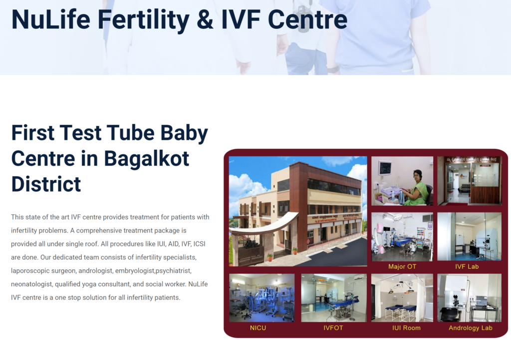
SpOvum is now adopted at NuLife Fertility & IVF Centre, Bagalkot

The SpOvum team is delighted to get started in yet another clinic in Bagalkot!

SpOvum is making strides in ensuring IVF hospitals excel in providing quality IVF treatment to the patients.

You can know more about the clinic here: http://www.manehospital.com/nulife.php

You can contact them here: http://www.manehospital.com/contact.php

The SpOvum team wishes the clinic a great success in the days to come, enabled by our SpOvum suite of products!

Bio-Insights-Embryo

Embryo Development

The product of fertilization is an embryo. The first stage is the one-cell embryo with a diploid number of chromosomes. The embryo undergoes a series of cell divisions, for few days to form a hollow sphere of cells known as blastocyst. [1]

Day-1, 2PN stage, in this stage, the embryo contains two polar bodies and two pronuclei with small nucleoli. One of the pronuclei contains genetic information from the egg and the other from the sperm. The pronuclei are usually checked between 16-18 hours after the sperm injection. [2]

The one cell embryo undergoes a series of cleavage divisions, progressing through 2-cell, 4-cell, 8-cell and 16-cell stages. The cells in cleavage stage embryos are known as blastomeres. Early on, cleavage divisions occur quite synchronously. In other words, both blastomeres in a two-cell undergo mitosis and cytokinesis almost simultaneously. Hence, due to these reasons, the embryos are most commonly observed at two, four and eight-cell stages. Embryos with an odd number of cells (e.g. 3,5,7) are less commonly observed, simply because those states last for a relatively short time. [1]

After the development of 8-cell embryo, the blastomere begins to form tight junctions with one another, leading to deformation of their round shape and formation of a mulberry-shaped mass of cells called a morula. This change in shape of the embryo is called compaction. It is difficult to count the cells in a morula, the embryo shown here probably has between 16 to 32 cells. [1 and 3]

Junctional complexes formed between the blastomeres gives the embryo outside and inside. The outer cells of the embryo express variety of membrane transport molecules, including sodium pumps, which results in an accumulation of fluid inside the embryo, which signals the formation of the blastocyst.

A blastocyst is composed of a hollow sphere of trophoblast cells, inside of which is a small cluster of cells called the inner cell mass. Trophoblast goes on to contribute to fetal membrane systems, while the inner cell mass is destined largely to become the embryo and fetus. Eventually, the stretched zona pellucida develops a crack and the blastocyst escapes by a process called hatching. This leaves an empty zona pellucida and a zona-free or hatched blastocyst lying in the lumen of the uterus. [1]

Embryo assessment

The most important morphological parameters to asses in the laboratory are blastomere appearance, fragmentation, and multinucleation. Those with equal blastomeres, minimal cytoplasmic fragmentation, and few multinucleated cells show a better prospect of implantation. [4]

  • Blastomere size

The relative blastomere size in the embryo is dependent on both the cleavage stage and the regularity of each cleavage division. [3]

  • Fragmentation

The fragmentation of the cytoplasm is therefore defined as the presence of anucleate structures of blastomeric origins. The degree of fragmentation is most often expressed as the percentage of the total cytoplasmic volume. The relative degree of fragmentation is defined as mild (<10%), moderate (10-25%) and severe (>25%). High degree of fragmentation correlates negatively with implantation and pregnancy rates, while the presence of minor amounts of fragmentation has no negative impact. [3]

  • Nucleation

The nucleation status is defined as the presence or absence of nuclei in the blastomeres of the cleavage stage embryo. Nucleation status of each blastomere in the embryo is evaluated as a single nucleus per blastomere, no nuclei visible or multinucleation. [3]. Multinucleation arises due to karyokinesis in the absence of cytokinesis with subsequent arrest of the blastomere or the entire embryo. Multinucleation may also be caused by partial fragmentation of nuclei or by defective migration at mitotic anaphase. [4] Multinucleation is predictive of a decreased implantation potential and are associated with an increased level of chromosomal abnormalities and increased risk of spontaneous abortion.


2PN embryo

The human oocyte arrested in the second meiotic division (M-II), usually characterized by the presence of a first polar body. The entry or injection of the sperm into the oocyte completes the second meiotic division. The second polar body containing the chromatids from one haploid chromosome set is extruded, and the female pronucleus is formed. During this process, the ooplasm rotates in a periodical way and in parallel the sperm chromatin decondenses. The sperm cell also delivers the centriole that has a leading role in further development and control of microtubules that are important for the symmetry of the developing embryo. These microtubules pull the haploid female pronucleus towards the male pronucleus. Both pronuclei finally migrate to the center of the cell and align. The G1 phase starts approximately 2-3 hr after sperm entry and pronuclei appear after 4-6 hr. The process is complete 18-22 hr after sperm entry or injection. [5]


Cleavage stage embryos

Embryos in cleavage stage range from the 2-cell stage to the compacted morula composed of 8-16 cells. Good quality embryos must exhibit appropriate kinetics and synchrony of division. In normal-developing embryos, cell division occurs every 18-20 h. Embryos diving either too slow or too fast may have metabolic and/or chromosomal defects. The blastomeres dividing in exact synchrony produces only 2, 4 or 8 cell embryos. Asynchronous developments lead to the formation of 3, 5, 6, 7 or 9 cell embryos. [3]

Mitosis in blastomeres should produce two equally sized daughter cells. When the division is asymmetric, one of the blastomeres of the next generation will inherit less than the amount of cytoplasm of parent blastomere, leading to a defective lineage in the embryo. After two cleavages, the zygote becomes a 4-cell embryo, where the 4-cells are normally arranged in a tetrahedron in the spherical space provided by the ZP. Blastomeres located close to a single, spatial plane produced by an incorrect orientation of division axes will lead to altered embryo polarity. [3]

After the embryo reaches the 8-cell stage, the blastomeres begin to show an increase in cell-cell adherence due to the spread of intercellular junctions. This is the start of compaction. The process of compaction advances during the next division until the boundaries between the cells are barely detectable. If some of the blastomeres are excluded from this compaction process, the embryo may have a reduced potential for becoming a normal blastocyst. [3]


Blastocyst

The blastocyst consists of cells forming an outer trophectoderm (TE/trophoblast) layer, an inner cell mass (ICM, embryoblast) and a blastocoel (fluid-filled cavity). The ICM forms an inner layer of larger cells also called as “embryoblast” which are the cluster of cells located and attached on one wall of the outer trophoblast layer. In week 2 of embryo development, ICM differentiates into two distinct layers the epiblast and hypoblast. ICM is the source of true embryonic stem cells capable of forming all cell types within the embryo. [7]

The trophectoderm (TE) outer layer of smaller cells is also called the “trophoblast” epithelium. The key function is for the transport of sodium (Na+) and chloride (Cl-) ions through this layer into the blastocoel. In week 2 this layer will differentiate into two distinct trophoblast layers the syncytiotrophoblast and cytotrophoblast cells and are key to implantation and early placentation. [7]


Blastocyst Hatching

As the fluid and number of cells inside the blastocyst increase its progressively causes enlargement of the blastocyst and its cavity with a consequent progressive thinning of the zona pellucida (ZP). Finally, the blastocyst breaks free of the ZP through a process called hatching.


Cytoplasmic Anomalies

The cytoplasm of cleaving embryos is normally pale, and clear or finely granular in appearance. Cytoplasmic anomalies, such as cytoplasmic granularity, cytoplasmic pitting and the presence of vacuoles, occur occasionally.

Cytoplasmic pitting is characterized by the presence of numerous small pits with an approximate diameter of 1.5 µm on the surface of the cytoplasm. The cytoplasm of blastomeres may be excessively darkened with centralized granularity, where these kinds of embryos have reduced implantation potential or can undergo degeneration.

Cytoplasmic vacuolization varies in size and number. Vacuoles are membrane-bound cytoplasmic inclusions filled with fluid that are virtually identical with the perivitelline fluid. Extensive vacuolization is always considered as detrimental. [6]

  1. http://www.vivo.colostate.edu/hbooks/pathphys/reprod/fert/cleavage.html
  2. https://ivf.net/ivf/embryo-development-o2591.html
  3. http://atlas.eshre.eu/es/14611830864735920
  4. The Infertility Manual, Editor: Kamini A Rao and Co-editor: Howard Carp
  5. https://books.google.co.in/books?id=Kp5_AwAAQBAJ&pg=PA111&lpg=PA111&dq=vacuoles+in+embryo+eshre&source=bl&ots=VCxpH1yiuz&sig=t1ARQo-CeYN24Isgl18iSY-3qeg&hl=en&sa=X&ved=2ahUKEwjBmqzNmsvcAhWCbn0KHTFfD3I4ChDoATABegQIAhAB#v=onepage&q&f=true (Morphological selection of gametes and embryos: 2PN/zygote by Martin Greuner and Markus Montag)
  6. http://atlas.eshre.eu/es/14611418225805670
  7. https://embryology.med.unsw.edu.au/embryology/index.php/Blastocyst_Development

Embryo Biopsy

  1. Blastomere Biopsy

Blastomere biopsy is a technique that is performed by removal of one or two cells (blastomeres) from 4-8 cell embryo for preimplantation analysis. On the third day of the embryo development, the embryo is maintained in a position by a pipette with rounded margins, an opening is made in the embryo by using a laser device or treating it with thyroid acid. Once the hole is made the cells from the embryo are removed using a micropipette having a greater diameter. At this stage of embryo development, all the cells are equivalent and thus removal of a cell from the embryo at this stage does not remove anything critical for normal development. The embryo compensates for the removed cell and should continue to divide following blastomere biopsy.  After removal of cells from the blastomere, the developing embryo is placed back into the culture dish and the removed cells are used for subsequent genetic analysis. [1]

2.  Trophectoderm Biopsy

Trophoblast/Trophectoderm are cells forming the outer layer of a blastocyst, which provide nutrient to the embryo and develop into a large part of the placenta. They are formed during the first stage of pregnancy and are the first cells to differentiate from the fertilized egg. [3] Trophectoderm biopsy involves removing some cells from the trophectoderm component of a blastocyst embryo at day 5/6.

Reference:

  1. http://www.preimplantationgeneticdiagnosis.it/polar-body-removal-blastomere-biopsy.htm
  2. https://nordicalagos.org/trophectoderm-biopsy/
  3. https://en.wikipedia.org/wiki/Trophoblast


Preimplantation Genetic Screening (PGS) and Preimplantation Genetic Diagnosis (PGD)

PGD: It was first introduced in the year 1989. A procedure to test the embryos for specific conditions before implantation in couples who are at risk of transmitting genetic abnormality to their offspring. The embryos are biopsied at either zygote, cleavage or blastocyst stage. Used to determine embryo genotype, performed in couples with genetic abnormalities such as single-gene disease, single mutations, translocations or other gene abnormalities. The embryos can be biopsied at different stages like zygote stage (removal of the first and second polar body), cleavage stage (removal of one to two blastomeres from the six to eight cell embryo) and blastocyst stage (removal of some trophectoderm cells). Almost all PGD cycles are carried out during blastomeres after the cleavage-stage biopsy. Polar body biopsy is rarely used as it only gives genetic information on the maternal genome. [1]

Testing by Fluorescence in-situ hybridization (FISH)

FISH involves identification of chromosomes or their fragments with fluorescently labeled molecular probes. Probes are complementary to specific DNA regions that are subject to hybridization under specific conditions, and the result of this process can be observed as fluorescent spots under a fluorescent microscope. In FISH it is not possible to test a whole panel of 24 chromosomes during one test, as it is only feasible to use 5-9 probes at most, for 2-3 rounds of hybridization. Hence, it is limited to the most common abnormalities involving chromosomes 13, 15-18, 21, 22, X and Y. Due to a lot of disadvantages of FISH, this field of study has now been successfully replaced by methods that are more reliable and precise, such as SNP and NGS. [2] This technique is usually referred to as preimplantation genetic screening (PGS) and should be differentiated from PGD, as it is for a different group of patients and for a different reason. [1]

Testing by PCR

For couples at risk of single gene disorder, PGD is carried out using PCR. [1]

Whole genome approaches

The introduction of whole genome amplification (WGA) methods has enabled high throughput technologies to be used, which has increased the amount and type of information that can be obtained from an embryo biopsy sample. Techniques that come under WGA are preimplantation haplotyping (PGH), Array comparative genome hybridization (CGH), Next-generation sequencing (NGS). These whole-genome approaches rely on whole-genome amplification. [1]

PGS: this is used to determine potential aneuploidies of all 24 chromosomes. Particularly performed for patients older than 35 (advanced maternal age), those with repeated implantation failures, repeated miscarriages, with severe male factor infertility.

Reference:

  1. https://www.rbmojournal.com/article/S1472-6483(15)00605-7/pdf
  2. https://www.researchgate.net/publication/306004985_Current_Methods_for_Preimplantation_Genetic_Diagnosis
 

Why is addressing infertility important?

Infertility is the inability to conceive after a year of trying or the inability to carry a pregnancy to term. Addressing infertility is important for several reasons:

  1. Fertility is an important part of reproductive health: Infertility can affect a person’s physical and mental health and can be a source of stress, anxiety, and depression. Addressing fertility issues can help improve overall reproductive health and well-being.
  2. It can improve the chances of having a successful pregnancy: There are various treatment options available for couples experiencing fertility issues, and addressing the underlying cause of infertility can increase the chances of having a successful pregnancy.
  3. It can help couples achieve their family-building goals: For many couples, starting a family is an important aspect of their lives. Addressing fertility issues can help couples achieve their goals of having children and building their families.
  4. It can help couples make informed decisions: Understanding the underlying cause of infertility can help couples make informed decisions about their treatment options and the potential risks and benefits of each option.

It is important for couples experiencing fertility issues to seek medical advice and support in order to address these issues and improve their chances of having a successful pregnancy.

DISCLAIMER: The information provided in here is for general educational purposes only and is not intended to be a substitute for professional medical advice, diagnosis, or treatment. Always consult a qualified healthcare provider for any questions you may have regarding a medical condition. Do not rely on the information provided here for decision-making or self-treatment. If you are experiencing a medical emergency, seek immediate medical attention.